Our research
Read more about our research interests below
Non-specialist synopsis
Leukaemia is a blood cancer that originates in the bone marrow through genetic mutations and results in the abnormal growth and survival of white blood cells. Acute myeloid leukaemia (AML) is a particularly aggressive form of this disease that affects both adults and children. Current chemotherapy drugs result in unacceptable levels of toxicity and disease relapse. New drugs that target specific molecular abnormalities in leukaemia cells are required in order to induce long-lasting remissions and reduce toxicity. One such molecular target which has emerged over the past decade is a protein called β-catenin which is the central player of the Wnt signalling pathway.
Signalling pathways such as Wnt are normally highly controlled mechanisms adopted by healthy cells to allow DNA modification in response to external signals. Very often, such mechanisms are hijacked by cancer cells to promote abnormal gene activation which in turns causes abnormal cell growth and survival (a hallmark of cancer). The level and activity of β-catenin is abnormally high in AML cells, which results in abnormal gene activation and confers inferior patient survival. In order for β-catenin to propagate its cancer-promoting effects it must first form partnerships with other molecules in the cell. Until now, β-catenin's repertoire of molecular partners in blood cells was unknown but our group has now discovered these partners in leukaemia cells for the first time.
Our research seeks to understand how the molecular partners of β-catenin boost its level and activity in leukaemia cells and investigate the biological significance of these partnerships for leukaemia progression. The ultimate aim of our research is to identify β-catenin molecular interactions of interest that could be exploited for therapeutic intervention and benefit AML patients.
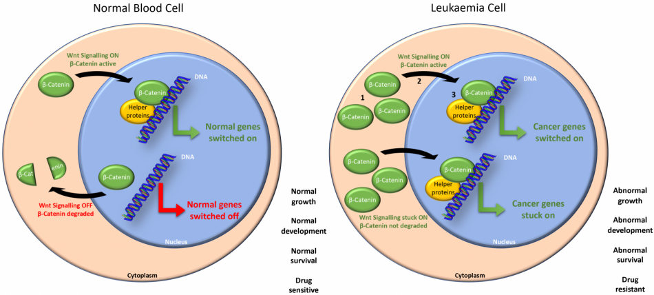
A simplified overview of how the cancer promoting protein β-catenin contributes to leukaemia progression. The lab is focussed on the following checkpoints numbered on the diagram:
1 - How does β-catenin protein accumulate in a leukaemia cell?
2 - How does β-catenin enter the nucleus of leukaemia cells?
3 - Which proteins bind β-catenin to increase its activity?
Specialist overview
The role of Wnt/β-catenin signalling in normal haematopoiesis and acute myeloid leukaemia (AML)
- The molecular events leading to aberrant stabilisation and activitation of β-catenin in AML
- The mechanisms regulating nuclear localisation of β-catenin in normal and malignant haematopoietic cells
- The role of Wnt signalling in normal myeloid development
- The characterisation β-catenin protein interactions in blood cells
- The development of novel therapeutic strategies for targetting Wnt signalling in haematological malignancy
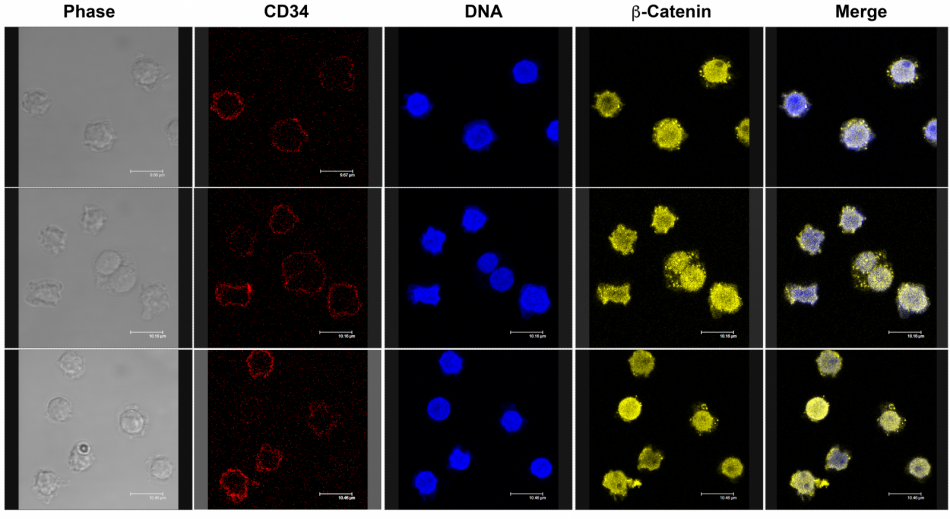
Localisation of the Wnt signalling protein β-catenin (yellow) in multiple fields of normal CD34+ (red) haematopoietic stem cells also showing phase contrast (grey), nuclear (blue) and merged images (Morgan RG et al, unpublished). Nuclear localisation of β-catenin is consistent with its role in the self-renewal of haematopoietic stem cells.
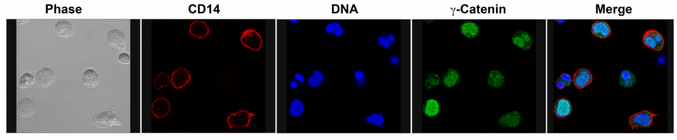
Localisation of the Wnt signalling protein γ-catenin (green) in normal CD14+ (red) monocytes also showing phase (grey), nuclear (blue) and merged images (Morgan RG et al, Leukaemia, 2013). Localisation of γ-catenin is highly variable within monocyte subpopulations.