Ligand effects on Quantum Dot Blinking
 Photoluminescent quantum dots are used in a range of applications that exploit the unique size tuneable emission, light harvesting and quantum efficient properties of these semiconductor nanocrystals. However, optical instabilities such as photoluminescence intermittency, the stochastic switching between bright, emitting states and dark states, can hinder quantum dot performance. Correlations between this blinking of emission and the dielectric properties of the nanoenvironment between the quantum dot interface and host medium, suggest surface ligands play a role in modulating on-off switching rates. Here we elucidate the nature of the cadmium selenide nanocrystal surface, by combining magic angle spinning NMR and x-ray photoelectron spectroscopy to determine ligand surface densities, with molecular dynamics simulation to assess net ligand filling at the nanocrystal interface. Results support a high ligand coverage and are consistent with photoluminescence intermittency measurements that indicate a dominant contribution from surface ligand to the dielectric properties of the local quantum dot environment. Go behind the science with Kudos
Sizing up excitons in Quantum Dots
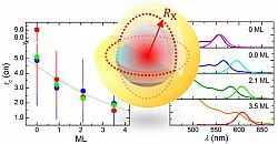 Semiconductor nanocrystals or quantum dots (QDs) are now widely used across solar cell, display, and bioimaging technologies. While advances in multishell, alloyed, and multinary core–shell QD structures have led to improved light-harvesting and photoluminescence (PL) properties of these nanomaterials, the effects that QD-capping have on the exciton dynamics that govern PL instabilities such as blinking in single-QDs is not well understood. We report experimental measurements of shell-size-dependent absorption and PL intermittency in CdSe-CdS QDs that are consistent with a modified charge-tunnelling, self-trapping (CTST) description of the exciton dynamics in these nanocrystals. By introducing an effective, core-exciton size, which accounts for delocalization of charge carriers across the QD core and shell, we show that the CTST models both the shell-depth-dependent red-shift of the QD band gap and changes in the on/off-state switching statistics that we observe in single-QD PL intensity trajectories. Further analysis of CdSe-ZnS QDs, shows how differences in shell structure and integrity affect the QD band gap and PL blinking within the CTST framework. https://pubs.acs.org/doi/10.1021/acsnano.7b01978
PCNA ubiquitylation influences PCNA chromatin association
PCNA ubiquitylation on lysine 164 is required for DNA damage tolerance. In many organisms PCNA is also ubiquitylated in unchallenged S phase but the significance of this has not been established. Using Schizosaccharomyces pombe, we demonstrate that lysine 164 ubiquitylation of PCNA contributes to efficient DNA replication in the absence of DNA damage. Loss of PCNA ubiquitylation manifests most strongly at late replicating regions and increases the frequency of replication gaps. We show that PCNA ubiquitylation increases the proportion of chromatin associated PCNA and the co-immunoprecipitation of Polymerase δ with PCNA during unperturbed replication and propose that ubiquitylation acts to prolong the chromatin association of these replication proteins to allow the efficient completion of Okazaki fragment synthesis by mediating gap filling. https://doi.org/10.1371/journal.pgen.1006789
Charge-tunnelling and self-trapping in Quantum Dots
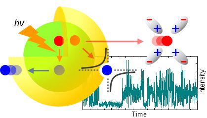 Understanding instabilities in the photoluminescence (PL) from light emitting materials is crucial to optimizing their performance for different applications. Semiconductor quantum dots (QDs) offer bright, size tunable emission, properties that are now being exploited in a broad range of developing technologies from displays and solar cells to biomaging and optical storage. However, instabilities such as photoluminescence intermittency, enhancement and bleaching of emission in these materials can be detrimental to their utility. Here, we report dielectric dependent blinking, intensity-“spikes” and low-level, “grey”-state emission, as well as PL enhancement in ZnS capped CdSe QDs; observations that we found consistent with a charge-tunnelling and self-trapping (CTST) description of exciton-dynamics on the QD–host system. In particular, modulation of PL in grey-states and PL enhancement are found to have a common origin in the equilibrium between exciton charge carrier core and surface-states within the CTST framework. Parameterized in terms of size and electrostatic properties of the QD and its nanoenvironment, the CTST offers predictive insight into exciton-dynamics in these nanomaterials. https://pubs.rsc.org/en/content/articlelanding/nr/2016/c6nr00529b
Single Molecule Localisation Microscopy of DNA-Associated Proteins

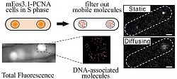
Development of single-molecule localization microscopy techniques has allowed nanometre scale localization accuracy inside cells, permitting the resolution of ultra-fine cell structure and the elucidation of crucial molecular mechanisms. Application of these methodologies to understanding processes underlying DNA replication and repair has been limited to defined in vitro biochemical analysis and prokaryotic cells. In order to expand these techniques to eukaryotic systems, we have further developed a photo-activated localization microscopybased method to directly visualize DNA-associated proteins in unfixed eukaryotic cells. We demonstrate that motion blurring of fluorescence due to protein diffusivity can be used to selectively image the DNA-bound population of proteins.We designed and tested a simple methodology and show that it can be used to detect changes in DNA binding of a replicative helicase subunit, Mcm4, and the replication sliding clamp, PCNA, between different stages of the cell cycle and between distinct genetic backgrounds. 10.1093/nar/gku726
DORIS : A Novel Super-Resolution Microscopy
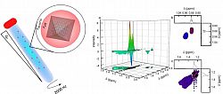
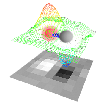 A novel super-resolution technique for resolving two or more fluorescent molecules, proximate within the diffraction-limit is developed. The method enables the identification of emitting centers that display intermittency in their fluorescence and separated by a distance in the 10-100 nm range, without the need for complex instrumentation or analysis. Direct object resolution by image subtraction (DORIS) uses the difference-image to resolve small but finite differences between two images of the PSF taken in time. The difference-image contains information on the relative orientation, separation and contribution of each fluorophore to the PSF in each frame. We exploit the fluorescence intermittency of QDs to show that, by subtracting pairs of normalized fluorescence images of dsDNA-coupled QDs, dimeric species can be readily visualised and distinguished from single QDs. Furthermore, we show that a simple relationship exists between the intensity amplitude in the difference-image of coupled-QDs and the distance between the centers of their respective PSFs. https://pubs.rsc.org/en/content/articlelanding/2013/CC/c3cc42072h
Sequencing by Synthesis (the Solexa way)
DNA sequence information underpins genetic research, enabling discoveries of important biological or medical benefit. Sequencing projects have traditionally used long (400-800 base pair) reads, but the existence of reference sequences for the human and many other genomes makes it possible to develop new, fast approaches to re-sequencing, whereby shorter reads are compared to a reference to identify intraspecies genetic variation. Here we report an approach that generates several billion bases of accurate nucleotide sequence per experiment at low cost. Single molecules of DNA are attached to a flat surface, amplified in situ and used as templates for synthetic sequencing with fluorescent reversible terminator deoxyribonucleotides. Images of the surface are analysed to generate high-quality sequence. We demonstrate application of this approach to human genome sequencing on flow-sorted X chromosomes and then scale the approach to determine the genome sequence of a male Yoruba from Ibadan, Nigeria. We build an accurate consensus sequence from >30x average depth of paired 35-base reads. We characterize four million single-nucleotide polymorphisms and four hundred thousand structural variants, many of which were previously unknown. Our approach is effective for accurate, rapid and economical whole-genome re-sequencing and many other biomedical applications. https://www.nature.com/articles/nature07517
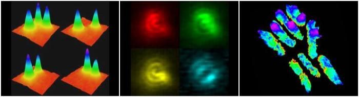
Single Molecule Dipole Imaging
 Single-molecule orientational imaging using total internal reflection fluorescence microscopy has been employed to investigate the dynamics of a protein−ligand system. Emission patterns from single tetramethylrhodamine (TMR)−biocytin molecules bound to streptavidin show that the TMR dipole adopts a limited number of favored orientations. The angular trajectories of individual dipoles exhibit remarkably similar patterns that are characteristic of single TMR molecules interacting with a relatively homogeneous population of nanoenvironments. Analysis of the polar and azimuthal angle distributions reveals a tendency for the dipole to assume three primary and two secondary orientations. Autocorrelation analysis of the dipole trajectories shows a predominantly bimodal behavior in the reorientation rates with the slow and fast components corresponding to the primary and secondary orientations, respectively. A number of mechanisms by which the observed orientations might be stabilized have been considered, in particular specific interactions between the zwitterionic TMR probe and charged residues on the streptavidin surface. Variations in the reorientation rates have been discussed in terms of local thermal fluctuations in the protein. https://pubs.acs.org/doi/10.1021/jp0517394
Single Molecule Spectroscopy of Semiconductor Nanocrystals
 The observation of photoluminescence (PL) activation, enhancement, fluorescence intermittency (FI), and decay over the photochemical lifetimes of single quantum dots (QD) is reported. The ability to count individual QDs along with PL intensities has revealed a measurable nonlinear correlation between the total fluorescence and QD population that is the result of enhancement and decay in the quantum yield (QY) of individual dots. This allows the differentiation between ensemble PL enhancement due to the fluorescence activation of a dark, non-emitting fraction of QDs and that due to genuine QY modification in individual nanocrystals. The evolution of the bright, fluorescent population under continuous illumination is seen to closely follow that of the intermediate in a consecutive elementary reactions scheme. Spectral separation of QD fluorescence trajectories into red and blue components indicates that PL decay is not necessarily associated with blue-shifting of the emission wavelength. https://pubs.acs.org/doi/full/10.1021/nn202994w
Single Molecule DNA Localisation Microscopy: towards Solexa (Ltd) SBS
 We have employed single molecule fluorescence spectroscopy, using a total internal reflection geometry and wide-angle detection, to study the attachment of singly fluorescently labeled DNA to a silica surface by either a streptavidin−biotin or a covalent linkage. In both cases the DNA is highly monodispersed with no evidence for aggregation. The covalent coupling gave higher signal-to-noise than the streptavidin−biotin linkage and was therefore studied in more detail. Two components in the photobleaching times, corresponding to different states of the tetramethyl rhodamine probe, were observed:
 a short and long component with populations in the ratio 6.7:1. Only rarely was interconversion between these two states detected during the 30-s observation time of the experiment. Hybridization experiments using a complementary strand of DNA labeled with a different fluorophore gave a low level of colocalized fluorescence, indicating a significant fraction of the surface attached DNA was not available for hybridization. These results are consistent with the surface attached DNA spending significant time collapsed on the surface. https://pubs.acs.org/doi/abs/10.1021/jp0038660