Preventing hearing loss due to treatment with aminoglycoside antibiotics
Aminoglycoside antibiotics enter hair cells in the inner ear through mechanosensitive ion channels (1), damaging the cells and eventually killing them (2,3). The cells can repair the damage provided exposure is brief (4): we aim to understand this repair process to see if it can be made more effective.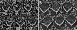
Damage to the surface of auditory hair cells following 15 minutes exposure to the aminoglycoside neomycin (A) is repaired after 15 minutes recovery without the drug (B). Picture by Richard Goodyear.
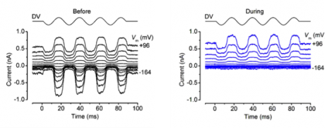
Patch-clamp recordings of sound-induced (mechano-electrical) currents in hair cells in cochlear cultures enable us to test novel compounds that could protect hair cells from aminoglycoside damage by blocking their entry pathway into the cells. Right hand panel shows voltage-dependent block with the peptide D-JNKi1.
Understanding the function of spontaneous electrical activity by inner hair cells before hearing onset
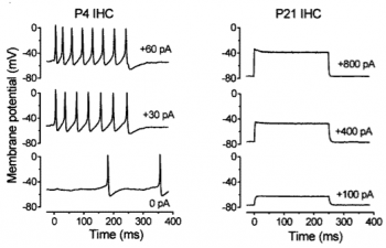
The function of cochlear inner hair cells is to signal the reception of sound to the brain. Before the onset of hearing, which occurs in mice about 12 days after birth (P12) the cells do not respond to sound, but instead fire spontaneous action potentials (left). After the onset of hearing the inner hair cells encode sound intensity by means of graded receptor potentials (right), so that louder sounds result in larger receptor potentials. From Kros (2007).
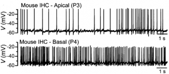
A clue to the function of the spontaneous action potentials comes from comparing their pattern along the cochlea. Immature inner hair cells in the apical coil of the cochlea (which will in the mature ear signal low-frequency sounds) fire action potentials in bursts (top). Action potentials of future high-frequency inner hair cells in the basal coil are random (bottom). This might be important for setting up appropriate tonotopic gradients (i.e. varying with sound frequency) along the auditory pathway in the brain and in the inner hair cells themselves. From Johnson et al (2011).
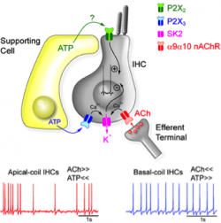
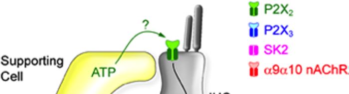
The differences in patterns of spontaneous action potentials between apical and basal inner hair cells may be due to difference in their modulation by acetylcholine (ACh) released by efferent nerve endings and by extracellular ATP released by neighbouring supporting cells. From Johnson et al (2011).