Medical Image Processing
Computed-Assisted Diagnosis based Texture Analysis (CAD-TA) for the predictive assessment and evaluation of medical images of cancer.
Current commercial CAD systems focus on automated detection of micro-calcifications and classification of masses as benign or malignant. CAD-TA has employed image feature quantification and artificial neural network based classifiers. The use of CAD to characterise pathological abnormalities and predict disease severity rather than detection is complex, more challenging and less developed. 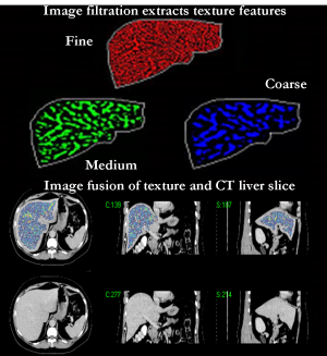
We have developed a novel CAD-TA method that differs from previous implementations of CAD-TA in two major aspects:
- Selective-scale feature filtration is employed for highlighting fine to coarse features and quantifying these extracted features separately and;
- Relative TA using texture ratio quantification is employed to assess the relative contributions made by features of different scales highlighted by the filter.
This technique has shown potential in colorectal and lung cancer on CT images and breast cancer on mammography, with possible applications to other organs obtained from different medical images such as MRI, PET, SPECT and Ultrasound etc.
We have also further developed 3-D texture analysis of whole volume which has shown potential in assessing pulmonary disorders such as pulmonary embolism and emphysema from Computed Tomography Pulmonary Angiography (CTPA) images of the lung.