High precision robotics and sensing for medical and industrial applications
We conduct research and development at the cutting-edge of high-precision robotics, advanced imaging, and sensing systems. We are interested in high precision robotics and its application in industrial and medical areas.
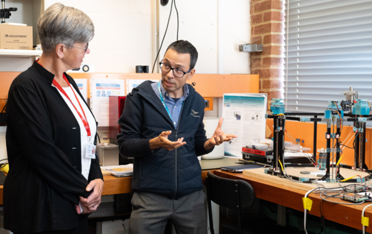
About the team
In the High Precision Robotics and Sensing Lab we design and develop the next generation high precision robotic systems combined with advanced imaging techniques and sensing capabilities. Our high precision robots are designed to be application specific achieving motion accuracies in the micrometre scale, enabling the development of future high precision medical and industrial applications.
Our areas of research expertise can be applied to a wide variety of applications such as:
- healthcare including robots and precision instruments for microsurgery and the inspection of medical devices and materials
- industrial manufacturing including the testing of products, devices and materials and defects identification
- quantitative imaging including the measurement of biomedical systems and processes through the extraction of objective numerical data from high resolution images
- pharmaceuticals including automated liquid handling, drug delivery kinetic, increasing precision and throughput in laboratory settings.
Consultancy services for businesses
We also provide dedicated expertise and undertake research on a consultancy basis to overcome complex technical challenges.
Find out more about our consultancy offering.
Research topics
Explore our research:
- Design and development of high precision robotic systems
- Ultrafast laser systems for microsurgery
- Advanced microscopy imaging techniques in robotics
- Photo-acoustic sensing for high precision multi-parametric quantification
- Additive manufacturing of stimuli responsive devices for biomedical applications.
Design and development of high precision robotics systems
Our goal is to improve human-assisted high precision tasks aiming to achieve an increasingly automated approach.
Our research focuses on improving robot accuracy to enable high precision tasks such as testing, micro-assembly and microsurgery.
We use experimental methods to develop and improve the precision of linear parallel robots covering from concept to fabrication. We develop application specific designs to achieve high repeatability considering the robotic system geometric structure, manufacturing tolerances and sensitivity to external disturbances. The use of multi-physical simulation and hardware design enabling us to address the robot structural and dynamic performance. Through calibration and control strategies we reduce the robot’s static and dynamic errors to enhance their accuracy, repeatability, and precision.
Our research also investigates pathways to enhance autonomy in our robotic systems by adopting image-based visual servoing (IBVS), position-based visual servoing (PBVS), and hybrid approaches, along with the development of robust algorithms for specific challenges such object identification, tracking and dealing with uncertainty in dynamic environments.
Applications: microsurgery, micro-assembly, high-throughput inspection, and precision testing.
- Academics
- References
[1] Huang, X.; Rendon-Morales, E.; Aviles-Espinosa, R. ROMI: Design and Experimental Evaluation of a Linear Delta Robotic System for High-Precision Applications. Machines 2023, 11, 1072. https://doi.org/10.3390/machines11121072
[2] Huang, X.; Rendon-Morales, E.; Aviles-Espinosa, R. Towards cellular level microsurgery: design and testing of a high precision delta robot for medical applications. Hamlyn Symposium on Medical Robotics 2023.
[3] Huang, X.; Rendon-Morales, E.; Aviles-Espinosa, R. Brightfield microscopy metrology system for the evaluation of high precision medical robotic system. 2023 Focus on Microscopy.
[4] Huang, X.; Rendon-Morales, E.; Aviles-Espinosa, R. A high precision robotic system design for microsurgical applications. 2023 MDPI Conference Engineering Proceedings
- Funders
- HEIF
- EPSRC
- Collaborators
-
University of Florence
-
Queen Victoria Hospital
- Royal Sussex County Hospital
-
Ultrafast laser systems for microsurgery
Our research focuses on the integration and evaluation of novel light sources with different wavelength beam and pulse characteristics for the next generation robotic microsurgical procedures.
We focus on understanding the light tissue interaction of ultrafast laser systems (including switched microchip, fibre and supercontinuum lasers) leading to the reduction of photo-damage for precise tissue removal. We use experimental photonic techniques including optical material characterisation, laser beam profiling and light transmission optimisation to adapt our technologies for microsurgical procedures with applications in tissue microdissection, cell biology and surface tissue characterisation.
We also conduct research with novel supercontinuum (white) lasers for medical applications including photodynamic therapies, tissue ablation and cauterization. By taking advantages of the properties of coherent light, we develop novel technologies to reduce tissue trauma and the invasiveness of contact based surgical methods, while reducing the time of tissue removal to improve patient's outcomes. We are one of the few research facilities in the UK with a supercontinuum laser system used for research.
- Academics
- References
[1] Huang, X.; Rendon-Morales, E.; Aviles-Espinosa, R. ROMI: A biomedical robotic platform combined with an application-specific laser-based end-effector for achieving high precision neurosurgery. IEEE Medical Measurements & Applications. IEEE proceedings 2024.
[2] Huang, X.; Rendon-Morales, E.; Aviles-Espinosa, R. Towards cellular level microsurgery: design and testing of a high precision delta robot for medical applications. Hamlyn Symposium on Medical Robotics 2023.
[3] Rodrigo Aviles-Espinosa, George Filippidis, Craig Hamilton, Graeme Malcolm, Kurt J. Weingarten, Thomas Südmeyer, Yohan Barbarin, Ursula Keller, Susana I.C.O Santos, David Artigas, and Pablo Loza-Alvarez, "Compact ultrafast semiconductor disk laser: targeting GFP based nonlinear applications in living organisms," Biomed. Opt. Express 2, 739-747 (2011)
- Funders
-
EPSRC
- HIEF
-
- Collaborators
-
Institute of Photonic Sciences (ICFO)
-
Technical University of Catalonia (UPC)
- Queen Victoria Hospital
-
Advanced robotic microscopy imaging techniques
Our research focusses on the integration of robotics with microscopy to create robotic microscopy systems, which enable automated, high-throughput, and precision imaging.
Our research also investigates the adoption of advanced light-based imaging techniques including brightfield, dark field, fluorescence, near-infrared (NIR), and selective plane illumination (SPIM) microscopy for quantitative imaging. By combining the in-house custom-made microscopes with wide working space robotic systems, we provide customised platforms that achieve precise sample manipulation and vision-guided systems for target tracking. We use multi-image stitching methods to achieve resolutions in exceeding 1.5 Giga pixels.
Our research combines novel AI and machine learning algorithms for image quantification and analysis including deep learning algorithms that automatically segment, register, and derive quantitative biomarkers/signature from images. This allows quantitative diagnostics with high resolution and contrast, faster image interpretation within a larger area of acquisition, identification of patterns, and data processing of complex datasets from robotic microscopy.
Applications: High-throughput screening, automated diagnostics in clinical settings, in vivo monitoring, advanced materials analysis and defect analysis of medical and industrial samples.
- Academics
- References
[1] Jiazhe Tang, X.; Aviles-Espinosa, R; Rendon-Morales, E. Machine vision system for quantification of aortic and pulmonic valvuloplasty catheter compliance. MPIE Conference 2024.
[2] Aviles-Espinosa, R; Dore, H; Williams, T; Rendon-Morales, E. Quantification of aortic valvuloplasty catheter size using a metrology system based on brightfield microscopy. European Focus on Microscopy Conference 2019.
[3] Rodrigo Aviles-Espinosa, G. Filippidis, Craig Hamilton, Graeme Malcolm, Kurt J. Weingarten, Thomas Südmeyer, Yohan Barbarin, Ursula Keller, David Artigas, and Pablo Loza-Alvarez "Compact ultrafast semiconductor disk laser for nonlinear imaging in living organisms", Proc. SPIE 7903, Multiphoton Microscopy in the Biomedical Sciences XI, 79032T (11 February 2011); https://doi.org/10.1117/12.874865
- Funders
- EPSRC
- HIEF
- Collaborators
- Institute of Photonic Sciences (ICFO)
- Technical University of Catalonia (UPC)
- Queen Victoria Hospital
- Sussex Cardiac Centre
- Royal Sussex County Hospital in Brighton
Additive manufacturing of stimuli responsive scaffolds for biomedical applications
Our research focuses on engineering innovation to design and test high precision stimuli responsive scaffolds.
We investigate the use of additive manufacturing (AM) technologies combined with advanced materials that have the ability to change their shape, properties and functions over time in response to external stimuli such as external magnetic fields and light sources.
Our research explores novel 3D printing methodologies for the manufacturing of magnetic scaffolds. We combine magnetic microparticles with polymers to create devices that can be experimentally guided to deliver drugs to specific sites in the body using external magnetic fields. Our techniques ensure uniform particle distribution, achieving high magnetic properties that are able of tailoring the material to the application's specific needs. We use multi-physical simulation to design complex structures and geometries that allow us to provide on-demand drug release.
We also study the interaction of the magnetic fields and the material properties including stiffness, shape and its relationship to drug release kinetics. We conduct experimental testing to assess the mechanical and structural properties of the scaffolds as well as the interacting magnetic fields allowing for high precision, targeted drug release for applications in precision medicine.
We also conduct research in the fabrication of novel light-stimuli scaffolds using photochromic biomaterials that respond to light, enabling precise control over cell behaviour, tissue development, and therapeutic delivery for applications in tissue engineering and regenerative medicine.
Applications: magnetic drug delivery systems (MDDS), target drug delivery, controlled drug released, tissue engineering, tissue regeneration and personalized medicine
- Academics
- References
[1] C. Yan, R. Aviles-Espinosa, S. Wang and E. Rendon-Morales, Design and Fabrication of 3D-Printed Magnetically Triggered Soft-Porous Scaffolds for Drug Delivery, 2024 International Conference on Networking, Sensing and Control (ICNSC), Hangzhou, China, 2024, pp. 1-5, doi: 10.1109/ICNSC62968.2024.10759996.
[2] C. Yan, R. Aviles-Espinosa, and E. Rendon-Morales, Towards the development of personalized drug delivery systems using 3D printed magnetically triggerable elastomers, 11th International Electronic Conference on Sensors and Applications, Engineering Proceedings, 82 (1) MDPI 2024
[2] Rendon-Morales E, Shi K, Woodbine L, Maniruzzaman M, Nokhodchi A, Aviles-Espinosa RA; Characterization of magnetically triggerable millirobots for on demand drug delivery using a brightfield microscopy metrology system. European Focus on Microscopy Conference 2022.
[3] Shi K, Aviles-Espinosa RA, Rendon-Morales E, Woodbine L, Salvage JP, Maniruzzaman M, Nokhodchi A, Magnetic field triggerable macroporous PDMS sponge loaded with an anticancer drug, 5-fluorouracil, ACS Biomaterials Science and Engineering7(1):180-195 American Chemical Society, 2021.
[4] Shi K, Aviles-Espinosa R, Rendon-Morales E, Woodbine L, Maniruzzaman M, Nokhodchi A, Novel 3D printed device with integrated macroscale magnetic field triggerable anti-cancer drug delivery system, Colloids and Surfaces B: Biointerfaces192 Elsevier 2020.
- Funders
- EPSRC
- HEIF
- Collaborators
-
University of Brighton
-
University of Mississippi
-
University of East London
-
Queen Victoria Hospital
-
Photo-acoustic sensing for high precision multi-parametric quantification
Our research focuses on engineering innovation to design and test novel photoacoustic sensors.
Our photoacoustic sensing technology is designed based on laser illumination, a principle used to generate self-interference random patterns. Through the analysis of the displacement and or temporal changes in such patterns when subjected to acoustic sources, high precision, remote, and non-contact measurements with sub-micron accuracies are possible.
We carry out research in speckle patterns engineering to ensure that the surface of the sample under inspection produces a varying intensity distribution with sufficient contrast, to identify the carrier of information such as deformation or vibration.
We combine our sensors with advanced imaging systems to analyse the generated speckle patterns that enables the extraction of information about a target's response to photoacoustic excitation.
We also conduct research on the application of machine learning approaches to process large and complex speckle datasets to achieve real-time parameters quantification for time-sensitive applications.
This results in the development of application-specific photo-acoustic sensors to tackle the challenges present in bio-medical diagnostics, including the measurement and multi-parametric quantification of micro-flows and the determination of biological samples composition, as well as for industrial applications including non-destructive testing, vibration, and motion monitoring.
Applications: Imaging including the monitoring micro flows in tissue, material vibrations, deformation, and non-destructive testing.
- Academics
- References
[1] Haji-Hasanli, H; Rendon-Morales, E; Aviles-Espinosa, R; Acousto-Optical Sensing of Tissue Composition in Laser-Assisted Robotic Microsurgeries. MPIE Conference 2025.
[2] Pablo Loza-Alvarez, David Artigas, and Rodrigo Aviles-Espinosa, “Semiconductor Lasers and Diode-based Light Sources for Biophotonics,” Chapter12, ISBN: 978-1-78561-272-5 the institute of engineering and technology IET 2018.
[3] P. Loza-Alvarez, R. Aviles-Espinosa, S.J. Matcher, D. Childs, S.G Sokolovski “Quantum Dot Ultrafast and Continuous Wavelength Laser Diodes for Applications in Biology and Medicine” in Book “The Physics and Engineering of Compact Quantum Dot-based Lasers for Biophotonics” Willey, Chapter 5, pp 171-230 (2013) ISBN: 978-3-527-66560-0
- Funders
- EPSRC
- HEIF
- Collaborators
-
University of Brighton
-
ICFO
- University of Florence
-