Broadcast: News items
Software improves cancer scans
By: James Hakner
Last updated: Tuesday, 26 January 2010
Thanks to a grant from the University's Enterprise Fund, researchers have developed new imaging software that highlights and quantifies previously invisible anomalies within cancer scans.
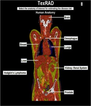
TexRAD can extract and quantify 'hidden' information from existing scans (CT, MRI, etc.), thereby increasing considerably the depth of information given by radiological images.
Professor Ken Miles of the Brighton and Sussex Medical School (BSMS), consultant radiologist and TexRAD clinical advisor, said: "TexRAD can assist clinical decision-making by predicting the risk of disease and assessing the prognosis for cancer patients."
TexRAD is an important development because it derives 'textures' from routine diagnostic images and highlights anomalies not apparent to the human eye. From these anomalies the software generates a risk-stratification report and can even be used retrospectively on old scan data.
TexRAD currently analyses CT images of colorectal, lung, renal and prostate cancers as well as mammography for breast cancer.
The Enterprise Fund provides funding and support to help staff get new products, solutions and creative commercial ideas off the ground. The money, channelled from the government's Higher Education Investment Fund, is used to encourage collaboration across campus to bring ground-breaking and enterprising ideas to fruition.